Concierge imaging · London
The right MRI, at the right London clinic - arranged by a clinician, not an algorithm.
Why patients choose us
- 01
A real person, every time
The same clinician stays with your enquiry from first message to final report.
- 02
Independent by design
We never accept payment to influence which clinic we recommend. Free for patients.
- 03
A vetted London panel
A small Harley Street network, re-vetted every six months on scanner quality and reporting.
Indicative pricing
Honest ranges, not "from £249" bait pricing.
Indicative ranges across our partner clinics. Send the details and we quote firm figures across two or three options.
In short
Most single-region MRIs in our network: £275-£500, reported in 24-72 hours.
| Scan type | Indicative range | Typical duration | Report turnaround |
|---|---|---|---|
| Single body part (knee, shoulder, brain, lumbar spine, etc.) | £275–£500 | 20–40 min | 24–72 hrs |
| Two body parts | £450–£800 | 40–60 min | 24–72 hrs |
| Three body parts | £650–£1,100 | 60–90 min | 24–72 hrs |
| Full spine (cervical, thoracic, lumbar) | £600–£950 | 45–75 min | 24–72 hrs |
| MRI with contrast (gadolinium) | + £50–£150 | +15 min | 24–72 hrs |
| Multi-parametric prostate MRI (mpMRI) | £550–£950 | 45–60 min | 48–72 hrs |
| Cardiac MRI | £750–£1,400 | 60–90 min | 48–72 hrs |
| Breast MRI | £600–£1,000 | 45–60 min | 48–72 hrs |
| Full-body MRI screening | £750–£1,900 | 60–90 min | 3–5 days |
| Paediatric MRI | Quoted case by case | Varies | 48–72 hrs |
| Open / upright MRI (claustrophobia-friendly) | £400–£700 | 30–60 min | 24–72 hrs |
Prices vary by clinic, time of day, scanner field strength (1.5T vs 3T), and whether contrast is used. We come back with a firm quote within one working day.
The problem
Booking an MRI in London is harder than it should be.
Prices vary fourfold. Half the clinics on Google are referral funnels. The wrong scan answers the wrong question. We vet the clinics, handle the booking and stay with you through the report - and when an MRI is not the right next step, we say so.
-
Not sure if you need an MRI?
We will say honestly - sometimes an ultrasound, X-ray or specialist consultation is the better first step.
-
Not sure which MRI?
Brain, spine, knee, prostate, cardiac - the right protocol matters as much as the right body part.
-
Not sure which clinic?
A small vetted network across central, north, west and south London - chosen on scanner quality and reporting.
The journey
From enquiry to report - what happens, in order.
One concierge, start to finish. Usually under a week from first message to consultant report.
Phase 1 · Before your scan
Concierge, off-stage for you
Phase 2 · On the day
~1 hour at the clinic
Phase 3 · After
Concierge, back on
- 01
Before
You tell us what is going on
A short, confidential form. Symptoms, timeline, referral or insurer if you have them.
- 02
Before
We come back with a recommendation
Within one working day: which scan, which clinic, indicative price. If MRI is not the right step, we say so.
- 03
Before
We arrange the appointment
Often same or next day, including evenings and Saturdays. Insurer pre-authorisation handled.
- 04
On the day
Arrival and safety check
Change into a gown, remove anything metallic. We have pre-completed the safety questionnaire.
- 05
On the day
In the scanner
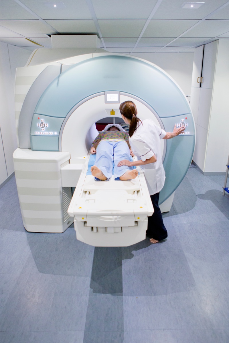
15-60 minutes. Loud, but with headphones, music and a two-way intercom throughout.
- 06
On the day
Straight home
No recovery time. Drive, eat and work as normal.
- 07
After
Report and next steps
Consultant radiologist report in 24-72 hours. We route it to your GP or specialist.
Typical end-to-end: 3-7 days. Urgent cases: same day.
By body part
Find the scan that matches the symptom.
Each guide covers when the scan is used, what it detects, price range and what to expect.
-
Brain & nerves
Headaches, dizziness, memory, suspected MS, stroke follow-up.
Learn more -
Spine
Back or neck pain, sciatica, slipped disc, post-op review.
Learn more -
Knee
Ligament tears, meniscal injury, cartilage, arthritis.
Learn more -
Shoulder
Rotator cuff, impingement, frozen shoulder.
Learn more -
Hip
Labral tears, impingement, avascular necrosis.
Learn more -
Ankle & foot
Sports injuries, tendinopathy, stress fractures.
Learn more -
Wrist & hand
Cartilage, ligaments, ganglia, post-traumatic.
Learn more -
Pelvis
General pelvic imaging, masses, post-op review.
Learn more -
Women's pelvic MRI
Endometriosis, fibroids, ovarian assessment.
Learn more -
Prostate (mpMRI)
The modern standard before any biopsy decision.
Learn more -
Abdomen
Liver, kidneys, pancreas, adrenals.
Learn more -
Cardiac MRI
Heart muscle, scarring, function.
Learn more -
Breast MRI
High-risk screening, treatment planning.
Learn more -
Full-body screening
A comprehensive look at major organs and spine.
Learn more
Specialist MRI types
Not all MRIs are the same.
What each option on your referral is actually for.
-
3T high-field MRI
Sharper images for brain, spine, prostate and small joints.
Read more -
1.5T MRI
Well-established and often preferred for patients with certain implants.
Read more -
Wide-bore MRI
A larger scanner opening for bigger or anxious patients.
Read more -
Open / upright MRI
For severe claustrophobia, or weight-bearing spine imaging.
Read more -
MRI with contrast
Extra characterisation for tumours, inflammation and vascular questions.
Read more -
Multi-parametric prostate MRI
The modern standard before any biopsy decision. Reported with PI-RADS.
Read more -
Cardiac MRI
Gold-standard non-invasive assessment of heart muscle and function.
Read more -
Paediatric MRI
Child-friendly clinics with experienced paediatric radiographers.
Read more
Our vetted London network
A small panel of clinics, we picked them.
Partners across central, north, west and south London. Not listed publicly - introductions are made privately, once we understand your case.
Selection criteria
How we choose every clinic in our network.

-
CQC-registered with a current good or outstanding rating
-
Reported by GMC-registered consultant radiologists, not generalists
-
Modern 3T or wide-bore 1.5T scanners
-
Report turnaround under 72 hours as standard
Safety and eligibility
One of the safest tests in medicine.
A few situations need specific checks. We handle them before you arrive.
-
Pacemakers, defibrillators, neurostimulators
Many modern devices are MR-conditional. We check the model before booking.
-
Cochlear implants
Case-by-case. We confirm with the device manufacturer.
-
Aneurysm clips and vascular coils
Model and date of insertion matter. We need this from your hospital.
-
Metal fragments (eye, shrapnel)
May need a prior orbit X-ray. We arrange this where required.
-
Joint replacements, stents, plates
Almost always fine. May cause local artefact but rarely a safety concern.
-
Dental work, tattoos, coils
Almost always fine. Iron-based tattoo ink may warm slightly - tell the radiographer.
-
Pregnancy
No ionising radiation. Generally safe, particularly after the first trimester. Contrast avoided unless essential.
-
Breastfeeding
Gadolinium is permitted. No need to pump and discard.
-
Kidney function
A recent eGFR is needed if contrast is planned.
-
Weight and size
Most scanners accommodate up to 200 kg. Wide-bore options for larger patients.
-
Children
Paediatric-friendly clinics. Sedation arranged where required.
Reading your report
A report can look intimidating. It isn't.
Almost every MRI report follows the same four-part structure.

A quiet reminder
Your report is written for your doctor, not for you.
If you would like us to talk you through it before your follow-up, just ask.
- 01 Header
Your details and the question
Who you are, what scan was performed, and the clinical question your referrer wanted answered.
- 02 Technique
How the scan was done
Which sequences were used (T1, T2, FLAIR, diffusion). Each highlights tissue differently.
- 03 Findings
What the radiologist saw
A region-by-region description, including normal anatomy and any incidental findings.
- 04 Impression
The conclusion: read this first
A short summary of what matters. Your specialist will discuss it in context.
Recognised by major UK insurers
Coverage varies by clinic and policy. We confirm with your insurer before booking.
Frequently asked
Everything we get asked before someone enquires.
Quick answers on referrals, insurance, claustrophobia and second opinions.
-
How quickly can I get an appointment?
Usually within one to three working days, including evenings and Saturdays. Same-day where clinically urgent.
-
Do I need a referral?
Most scans accept self-referral. Cardiac, paediatric and mpMRI prostate need a letter - we can arrange a fast-track private GP if needed.
-
Will my insurance cover this?
Most policies cover MRI when medically indicated and pre-authorised. Bupa, AXA, Vitality, Aviva, WPA, Cigna and Healix are recognised. We handle pre-authorisation.
-
Can I have an open MRI for claustrophobia?
Yes. Several partner clinics offer wide-bore, open or upright MRI. Tell us and we will route you there.
-
What's the difference between 1.5T and 3T?
3T gives sharper images for brain, spine, prostate and small joints. 1.5T is well-established and often preferred with certain implants. We match to your case.
-
Will I get the images, not just the report?
Yes. You get the consultant radiologist report and the full DICOM image set on USB or secure online access.
-
Can the report go to my GP or consultant?
Yes. With your consent, we route the report and images directly to your referring clinician the day they are signed off.
-
Can I have a full-body MRI screening without symptoms?
Yes. It has real value and real limitations, particularly the rate of incidental findings. We explain both before you book.
-
Are children scanned at your clinics?
Yes. Paediatric-friendly clinics with experienced radiographers. Sedation arranged by a consultant paediatric anaesthetist where required.
-
What does a second-opinion read cost?
Typically £250-£450 depending on complexity. We arrange this with a reporting consultant - no need to re-scan.
-
Will the MRI hurt?
No. Painless. Loud, so you have headphones or earplugs, and you need to lie still.
-
Can I have an MRI if I'm claustrophobic?
Yes. Open-bore scanners, music, eye masks, a companion, or a mild sedative if needed. Tell us and we will match you to the right clinic.